
Investigators have uncovered mind alterations in patients up to six months after they recovered from COVID-19.
Scientists uncovered brain changes in patients up to 6 months after they recovered from COVID-19 through the use of a different classification of MRI. here's in keeping with a examine that might be presented at the annual meeting of the Radiological Society of North the us (RSNA) next week.
in accordance with the U.S. facilities for disorder manage and Prevention (CDC), approximately one in five adults will strengthen lengthy-time period effects from COVID-19. issue thinking or concentrating, sleep complications, headache, lightheadedness, trade in smell or taste, pins-and-needles sensation, and depression or anxiousness are all neurological symptoms linked to long COVID. however, analysis reports have found that COVID-19 can be linked to adjustments to the coronary heart, lungs, or other organs even in asymptomatic sufferers.
As extra individuals become infected and recuperate from COVID-19, analysis has begun to emerge, focusing on the lasting consequences of the sickness. These are known as publish-COVID circumstances, which might be also wide-spread with the aid of a myriad of names including long COVID, lengthy-haul COVID, submit-acute COVID-19, publish-acute sequelae of SARS CoV-2 infection (PASC), long-time period consequences of COVID, and chronic COVID.
For this analyze, researchers used susceptibility-weighted imaging to analyze the outcomes that COVID-19 has on the brain. Magnetic susceptibility denotes how a great deal definite materials, equivalent to blood, iron, and calcium, will develop into magnetized in an applied magnetic box. This skill aids within the detection and monitoring of a host of neurologic circumstances including microbleeds, vascular malformations, mind tumors, and stroke.
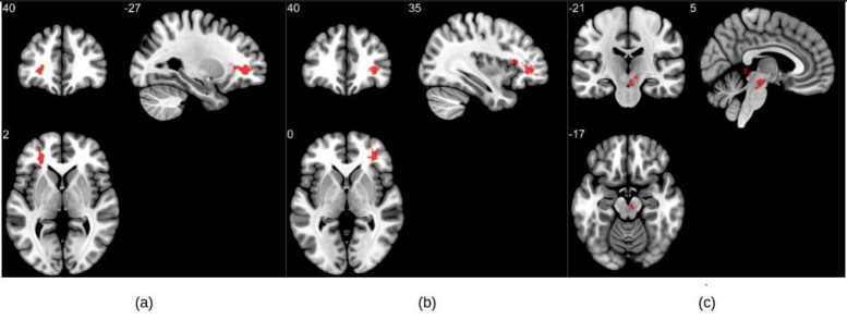
community analysis on susceptibility-weighted imaging exhibiting higher susceptibility-weighted imaging values within the COVID community when in comparison to suit controls. Three massive clusters have been found basically within the white be counted regions of the pre-frontal cortex and in the brainstem. The clusters (a) and (b) are followed bilaterally within the cerebral white count number close the orbitofrontal gyrus, whereas (c) lies in the midbrain location. credit score: RSNA and Sapna S. Mishra
"neighborhood-degree experiences haven't up to now focused on COVID-19 adjustments in magnetic susceptibility of the mind despite several case reports signaling such abnormalities," stated analyze co-writer Sapna S. Mishra, a Ph.D. candidate on the Indian Institute of know-how in Delhi. "Our analyze highlights this new factor of the neurological effects of COVID-19 and reviews giant abnormalities in COVID survivors."
The researchers analyzed the susceptibility-weighted imaging information of 46 COVID-recovered sufferers and 30 fit controls. Imaging become accomplished inside six months of restoration. amongst sufferers with long COVID, the most commonly suggested indicators were fatigue, problem dozing, lack of consideration, and reminiscence issues.
"alterations in susceptibility values of brain regions could be indicative of native compositional alterations," Mishra talked about. "Susceptibilities may additionally replicate the presence of irregular quantities of paramagnetic compounds, whereas lessen susceptibility may be brought about through abnormalities like calcification or lack of paramagnetic molecules containing iron."
MRI results confirmed that sufferers who recovered from COVID-19 had tremendously higher susceptibility values in the frontal lobe and mind stem compared to fit controls. The clusters received within the frontal lobe basically show variations within the white count number.
"These brain regions are linked with fatigue, insomnia, anxiousness, depression, headaches, and cognitive issues," Mishra noted.
portions of the left orbital-inferior frontal gyrus (a key place for language comprehension and creation) and appropriate orbital-inferior frontal gyrus (linked to numerous cognitive functions including attention, motor inhibition, and imagery, in addition to social cognitive methods) and the adjoining white rely areas made up the frontal lobe clusters.
The researchers also found a major difference in the right ventral diencephalon place of the brain stem. This place is associated with many essential bodily functions, including coordinating with the endocrine equipment to release hormones, relaying sensory and motor indicators to the cerebral cortex and regulating circadian rhythms (the sleep-wake cycle).
"This analyze aspects to critical lengthy-term complications that could be brought about with the aid of the coronavirus, even months after restoration from the infection," Mishra noted. "The existing findings are from the small temporal window. besides the fact that children, the longitudinal time facets throughout a few years will elucidate if there exists any permanent alternate."
The researchers are conducting a longitudinal study on the same affected person cohort to determine no matter if these mind abnormalities persist over a longer time body.
Co-authors are Rakibul Hafiz, Ph.D., Tapan Gandhi, Ph.D., Vidur Mahajan, M.B.B.S., Alok Prasad, M.D., and Bharat Biswal, Ph.D.
assembly: 108th Scientific meeting and Annual assembly of the Radiological Society of North the us
Post a Comment